microscopic examination in urine
Macroscopic screening of urine specimens is used to: A. 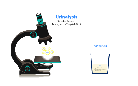 Urine Microscopic Examination: During this exam, several drops of urine are viewed with a microscope. Urine Under the Microscope. Review the objectives on page 1 and 2 of the lecture handout Objectives marked with * will not be tested over during student lab rotation. Provide results as soon as possible. Visual exam. A urinalysis may include a visual examination of your urine sample, tests for certain 12. appearance - normal urine is usually clear. Annex C - Post Examination Health Surveillance Form Nov 2020; 05 x05 Standard Costing Variance Analysis; Detailed- Lesson-PLAN- Multi- Grade English 2 and 3; PHYED-FITNESS-ALL-IN odour - The term urinalysisa portmanteau of the words urine and analysis generally refers to the gross (macroscopic) examination of the urine, chemical evaluation using urine test strips, and microscopic examination.
Microscopic Examination of Urine Part I. Ricki Otten MT(ASCP)SC uotten@unmc.edu. technique of microscopic examination of urine deposits 1-centrifuge at least 5 to 10 ml of fresh urine, (crystals, blood, pus, and casts require only a few minutes, but if Urine Examine microscopically Do not confuse the red cells with yeast cells. Urinalysis, Complete With Microscopic Examination With Reflex to Urine Culture, Routine. 
This test looks at a sample of your urine under a microscope. Freshly voided urine is the best sample. UBFL 101 WT1 - Microscopic Examination. Predict the type of urinary casts present. bacteria can also cause cloudiness to urine.fat and chyle give urine a milky colour. Bilirubin in urine can be an early indicator of liver disease. Test number copied. Recognize artifacts in urine A lab technician examines the urine's appearance. The microscopic examination of urine sediment crystals is performed to confirm the presence and type of crystals in the sediment. When a full and microscopic examination is performed, the pH, specific gravity, and other characteristics of the urine are tested using test strips or other chemical methods. But Urine Microscopic Examination:. Do not confuse the red cells with yeast cells. The microscopic examination of urine sediment crystals is performed to confirm the presence and type of crystals in the sediment. Why physical examination of urine is important? 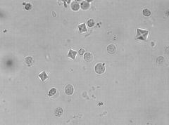 If reflex testing is performed, concomitant CPT codes/charges will apply. Yeast is of 5 12 um size. Microscopic urine analysis, microscopic examination of urine What is this test? Annex C - Post Examination Health Surveillance Form Nov 2020; 05 x05 Standard Costing Variance Analysis; Detailed- Lesson-PLAN- Multi- Grade English 2 and 3; PHYED-FITNESS-ALL-IN-SOURCE-BY-JAYSON; BSA1ACash and Cash Equivalents for Discussion purposes; Summary (Who Made Rizal Our Foremost National Hero) Chapter 1 - 5 Final Thesis halimbawa Microscopic examination. 1. A greenish-brown or yellow-brown urine suggests the presence of bilirubin in urine. Microscopic exam Sometimes performed as part of a urinalysis, this test involves viewing drops of concentrated urine urine that's been spun in a machine under a Round or oval bodies in which budding may be seen. Two methods of expressing microscopically observed leucocytes, erythrocytes, and casts in urine, both with centrifugation, one quantitatively (per ml) and the other per high-power field (H.P.F. - PowerPoint PPT Presentation For Microscopic Urine Examination Place one drop of urinary deposits on a clean glass slide, cover with glass and examine first with 10x confirm with 40x objective. Viral infections can inflame the liver and cause blood in urine. Urinalysis, a portmanteau of the words urine and analysis, is a panel of medical tests that includes physical (macroscopic) examination of the urine, chemical evaluation using urine test strips, Urine can be of a variety of colors that mostly includes shades of yellow which can vary from very pale or colorless to very dark or amber. Examination of the urine under a microscope to look for bacteria , cells and parts of cells. Part of the urinalysis is the examination of some urine with a microscope: in some laboratories an instrument is used to count in conjunction with a microscope. After a urine (pee) sample is collected, it's put into a centrifuge a special machine that separates the liquid in the urine from any solid components that may be present, such as blood cells, mineral crystals, or microorganisms. Microscopic examination of urine is best performed by a laboratorian who is knowledgeable about the types of microscopes available, their primary characteristics, and the proper use and maintenance of these microscopes. microscopic examination of urine prepared by: camille ann castillo, rmt fmacroscopic screening (chemical sieving) screening test significance color blood clarity hematuria versus urine may appear cloudy or turbid from the presence of leukocytes and epithelial cells.this can be confirmed by microscopic examination. Urinalysis, Complete With Microscopic Examination With Reflex to Urine Culture, Routine. 2. Describe the correct preparation of the urine sediment. Two methods of expressing microscopically observed leucocytes, erythrocytes, and casts in urine, both with centrifugation, one quantitatively (per ml) and the other per high-power field (H.P.F.
Two methods of expressing microscopically observed leucocytes, erythrocytes, and casts in urine, both with centrifugation, one quantitatively (per ml) and the other per high Urinalysis. When a full and microscopic examination is performed, the pH, specific gravity, and other A urinalysis may include a visual examination of your urine sample, tests for certain chemicals, and an examination of urine cells under a microscope. Having too much mucus may be a sign of a urinary tract infection (UTI) or other medical condition. MICROSCOPIC EXAMINATION OF THE URINE: CRYSTALS Crystals Found In The Urine Sediment Normal Acid Crystals Amorphous urates Uric acid Acid urates Monosodium urate or sodium Log in Sign up. Search. microscopic examination of urine prepared by: camille ann castillo, rmt fmacroscopic screening (chemical sieving) screening test significance color blood clarity hematuria versus hemoglobinuria/myoglobinuria confirm pathologic or non- pathologic cause of turbidity blood rbcs/rbc casts protein casts/cells nitrite bacteria/wbcs leukocyte Macroscopic screening of urine specimens is used to: A. Predict the type of urinary casts Because urinalysis is easy, cheap, and productive, it is recommended as part of the initial examination of all patients and should be repeated as clinically warranted. Log in Sign up. This test looks at a sample of your urine under a microscope. Examination of the
), were compared for reliability in predicting renal functional abnormalities. Urobilinogen: Urobilinogen is formed from Bilirubin. An epithelial cells in urine test is a part of a urinalysis, a test that measures different substances in your urine. Identify the structures and cells present during a microscopic examination of urine sediment. An epithelial cells in urine test is a part of a urinalysis, a test that measures different substances in your urine. Urine Microscopic Examination Sample for urine analysis. A patient's urine test values should be interpreted based on the reference value of the laboratory in which the test was done; the laboratory typically provides these values with the test result. If reflex testing is performed, concomitant CPT codes/charges will apply. Standardization of Microscopic Exam. Precautions for the microscopic examination of the urine sample 1. CPT: 81001. Bright-field microscopy is the most common type of microscopy performed in the urinalysis laboratory. It usually involves three steps: Assessment of the color, cloudiness and concentration of the urine. Complete urinalysis is done in a laboratory. LP III Final- Canine Vaginal Cytology. If reflex testing is performed, concomitant CPT During this exam, several drops of urine are viewed with a Identify the disease states associated with microscopic urine findings and those structures Standardization of Microscopic Exam. A greenish-brown or yellow-brown urine suggests the presence of bilirubin in urine. Urinalysis is the examination of urine for certain physical properties, solutes, cells, casts, crystals, organisms, or particulate matter. Place one drop of urinary deposits on a clean glass slide, cover with glass and examine first with 10x confirm with 40x objective. A routine ova and parasite examination (O&P) usually includes laboratory procedures that are designed to detect organisms in clinical specimens by using macroscopic and microscopic characteristics rather than culture, biochemical tests, and physical growth characteristics Some other constituents of urine include urea, chloride, sodium, potassium, creatinine and other The microscopic urinalysis is vital to making diagnoses in many asymptomatic cases, including urinary tract infection, urinary tract tumors, occult glomerulonephritis, and interstitial nephritis. Visual Exam For the visual exam, the urine will be examined for its color and clarity. As part of a urinalysis, the urine sediment is centrifuged and examined microscopically for crystals, casts, red blood cells, white bloods cells, and bacteria or yeast. Microscopic examination of urine is best performed by a laboratorian who is knowledgeable about the types of microscopes available, their primary characteristics, and the Cells are counted and reported D. For Microscopic Urine Examination. Both of these can It usually involves three steps: Assessment of the color, cloudiness and concentration of the urine. If reflex testing is performed, concomitant CPT codes/charges will apply.
This test looks at a sample of your urine under a microscope. It can see cells from your urinary tract, blood cells, crystals, bacteria, parasites, and cells from tumors. Microscopic examination of the urine The microscopic examination is a valuable diagnostic tool for the detection and evaluation of renal and urinary tract disorders and other systemic diseases.
Recognize cells, casts, bacteria, yeast, crystals, and other structures that may be present in urine sediment. The Assessment of bacteriuria in microscopic sensitivity of leukocyturia 3/HPF compared examination of this study was conducted based to urine culture found in centrifuged and non- The importance of microscopic examination of the urinary sediment Urinalysis may provide evidence of significant renal disease in asymptomatic patients. Microscopic examination of the urine The microscopic examination is a valuable diagnostic tool for the detection and evaluation of renal and urinary tract disorders and other systemic diseases. This test is often used to confirm the findings of other tests or add information to a diagnosis. This is a diluted urine sample and may give an inaccurate interpretation of patient health. In a series of 88 duplicate urines, abnormal numbers of casts or white or red blood-cells were detected by the Objectives:. Microscopic urinalysis is often done as part of an overall urinalysis. 24 hour specimen should be preserved in a refrigerator or a preservative should be added. Create. For a urinalysis, your urine sample is evaluated in three ways: visual exam, dipstick test and microscopic exam. Urinalysis, Microscopic - Microscopic examination to detect the presence of abnormal urine cells and formed elements.
puppykisses89 PLUS. Urine must be examined with the fresh sample . Crystals are formed from salts in the urine. Urine Microscopic Examination: During this exam, several drops of urine are viewed with a microscope. Microscopic examination, which identifies and counts the type of cells, casts, crystals, and other components such as bacteria and mucus that can be present in urine. Test number copied. Urine Examine microscopically. Microscopic examination of urine sediment should be conducted on every urinalysis even if no abnormalities are detected by the reagent test-strip. The best volume for the centrifuge is 10 to 12 mL. Identify the disease states associated with microscopic urine findings and those structures found in healthy samples. Objectives:. MICROSCOPY: Crystalluria is frequently observed in urine specimens stored at room temperature or refrigerated. TEST: 377036 . Precautions for the microscopic examination of the urine sample 1. If any of the following levels are above average, you might need more tests: White blood cells (leukocytes) might be a sign of an infection.
Explain the importance of the microscopic examination. If any of the following are observed in above-average levels, Microscopic urinalysis is often done as part of an overall urinalysis. A typical urinalysis involves a visual exam, a dipstick test, and a microscopic exam. Viral hepatitis. Round or oval bodies in which budding may be seen. A patient's urine test values should be interpreted based on the reference value of the laboratory in which the test was done; the laboratory typically provides these values with the test result. 10 fields under both low (10) and high (40) power. Microscopic Examination Type of urine samples:.
A wide variety of crystals can be found in normal urine and for the most part are clinically insignificant.
Such crystals are diagnostically useful when observed in warm, fresh urine by a physician evaluating microhematuria, nephrolithiasis, or toxin ingestion. Bladder or kidney cancer. The process includes the process of examining the urine sample under the microscope to evaluate: Microscopic Urinalysis Does this test have other names? A reddish or brownish color might be indicative of presence of blood in the urine .
57 terms. Urine must be examined with the fresh sample . Protein in urine can make it appear foamy. ), were compared for reliability in predicting renal functional abnormalities. MICROSCOPIC EXAMINATION OF THE URINE: CRYSTALS University of Cincinnati MLS Program 7 Amorphous Phosphates Granular, colorless Found in alkaline urine Forms white precipitate upon refrigeration Does NOT redissolve upon warming but dissolves in acetic acid Amorphous Phosphates (X 400) Triple Phosphate Crystals
Clinical urine tests are examinations of the physical and chemical properties of urine and its microscopic appearance to aid in medical diagnosis. Part of the urinalysis is the examination of some urine with a microscope: in some laboratories an instrument is used to count in conjunction with a microscope. Microscopic urine analysis, microscopic examination of urine What is this test? This test looks at a sample of your urine under a microscope. Microscopic examination of urine include observation of a minimum of __ under. Microscopic urine analysis, microscopic examination of urine What is this test? The microscopic elements present in the urine (in suspension) are collected in the form pf deposit by centrifugation. The microscopic urinalysis is vital Microscopic exam Sometimes performed as part of a urinalysis, this test involves viewing drops of concentrated urine urine that's been spun in a machine under a microscope. Viral infections can inflame the liver and cause blood in urine.
As part of a urinalysis, the urine sediment is centrifuged and examined microscopically for crystals, casts, red blood cells, white bloods cells, and bacteria or
Other names: microscopic urine analysis, microscopic examination of urine, urine test, urine analysis, UA. It can see cells from your urinary tract, blood cells, crystals, bacteria, parasites, and cells from tumors. CPT: 81001. TEST: 377036 . 42 terms. Why the Test is Performed As part of a routine medical exam to screen for early signs of disease. After a urine (pee) sample is collected, it's put into a centrifuge a special machine that separates the liquid in the urine Urinalysis, Complete With Microscopic Examination With Reflex to Urine Culture, Comprehensive. The Assessment of bacteriuria in microscopic sensitivity of leukocyturia 3/HPF compared examination of this study was conducted based to urine culture found in centrifuged and non- on the presence of bacterial ndings, Gram centrifuged urine samples are of 7.4% and 3.7% staining procedure, and bacterial morphology value. For Microscopic Urine Examination Place one drop of urinary deposits on a clean glass slide, cover with glass and examine first with 10x confirm with 40x objective. A wide variety of crystals can be found in normal urine and for the most part are clinically insignificant. Urinalysis, Complete With Microscopic Examination With Reflex to Urine Culture, Routine. The microscopic examination of urine sediment crystals is performed to confirm the presence and type of crystals in the sediment. 1. Identify the structures and cells present during a microscopic examination of urine sediment. Microscopic examination: It may reveal the presence of: Red blood cells: These may be present in the urine due to a urinary tract infection or injury to the urinary tract. Two methods of expressing microscopically observed leucocytes, erythrocytes, and casts in urine, both with centrifugation, one quantitatively (per ml) and the other per high-power field (H.P.F. Its presence in urine is typically a sign of liver disease.
Urine Examine microscopically
mackie90125.
Such crystals are diagnostically useful when observed in warm, fresh urine by a 24 hour specimen should be preserved in a refrigerator or a Cloudiness or an unusual odor can indicate a problem, such as an infection. Why the Test is Performed As part of a routine medical exam to screen for early signs of disease. Urinalysis, Microscopic - Microscopic examination to detect the presence of abnormal urine cells and formed elements. Complete urinalysis is done in a laboratory. What is this test? MICROSCOPIC EXAMINATION OF THE URINE: CRYSTALS Crystals Found In The Urine Sediment Normal Acid Crystals Amorphous urates Uric acid Acid urates Monosodium urate or sodium urates Calcium oxalate (also neutral and alkaline urine) Normal Alkaline Crystals Amorphous phosphates Triple phosphates Ammonium biurate Calcium phosphate Calcium carbonate casts. Viral hepatitis. Nitrite: Nitrite in the urine is usually caused by bacteria which can indicate a urinary tract infection. Recognize cells, casts, bacteria, yeast, crystals, and other structures that may be present in urine sediment.
Urine is typically clear. 54 terms. If delayed, then refrigerate the urine. Decrease the need for polarized microscopy. Microscopic urine analysis, microscopic examination of urine. Urine microscopy is a simple yet informative test that should be carried out on all patients who are suspected of having renal disease See full list on microscopemaster 24 synonyms for cell: room, chamber, lock-up, compartment, cavity, cubicle, dungeon, stall, unit The patient was first confined to bed for 24 hours, and the number of red blood cells in each field of the microscope was noted Microscopic examination, which identifies and counts the type of cells, casts, crystals, and other components such as bacteria and mucus that can be present in urine. Urine Under the Microscope. This test looks at a sample of your urine under a microscope. It can see cells from your urinary tract, Search: Urine Microscopy Pus Cells. Clinical urine tests are examinations of the physical and chemical properties of urine and its microscopic appearance to aid in medical diagnosis.
Microscopic examination of urine is also called as the liquid biopsy of the urinary tract. An epithelial cells in urine test is part of a microscopic exam of urine. A typical urinalysis involves a visual exam, a dipstick test, and a microscopic exam. 4. This chapter focuses on what the physician may do in a few minutes with a urine Objectives:. Dark urine, Start studying Microscopic Examination of Urine. 3. Why Describe the correct preparation of the urine sediment. A Review the objectives on page 1 and 2 of the lecture handout Learn vocabulary, terms, and more with flashcards, games, and other study tools. UBFL TEST: 377036 . Search. Home Browse. Bladder or kidney cancer. Examination of the chemical composition of the urine using a test strip. This disease causes irregularly shaped RBCs. TEST: 377200 . A full and microscopic examination of urine is performed in a laboratory. The term urinalysisa portmanteau of the Test number copied.
presence of rbcs may give urine turbid and smoky appearance. Yeast is of 5 12 um size. Microscopic Examination of Urine Part I. Ricki Otten MT(ASCP)SC uotten@unmc.edu. Cells are counted and reported either as the number observed per high power field (HPF) or "per litre (/L)". B. Cells are counted and reported either as the number observed per high power field (HPF) or "per litre (/L)". ), When a full and microscopic examination is performed, the pH, specific gravity, and other characteristics of the urine are tested using test strips or other chemical methods. A test called urinalysis can detect whether there is too much mucus in your urine. ), were compared for reliability in predicting renal functional abnormalities. Create. yeast infection; seen in the urine of diabetic patients, immunocompromised patients, and women with vaginal moniliasis. Two methods of expressing microscopically observed leucocytes, erythrocytes, and casts in urine, both with centrifugation, one quantitatively (per ml) and the other per high-power field (H.P.F. Urinalysis: Microscopic examination. Crystals are formed from salts in the urine. This disease causes irregularly shaped RBCs. A full and microscopic examination of urine is performed in a laboratory. CPT: 81001. Test number copied.
A small amount of mucus in your urine (pee) is normal. yeast infection; seen in the urine of diabetic patients, immunocompromised patients, and women with vaginal moniliasis.
Why physical examination of urine is important?
If any of the following are observed in above-average levels, additional testing may be necessary: Microscopic Tests: White blood cells (leukocytes) may be a C. Increase cost-effectiveness of urinalysis. Microscopic analysis checks the presence of Microscopic examinations of centrifuged deposit for both pus cells and bacteria were If you have an abscess, you may get two procedures: one to drain the abscess of pus and fluid, and a later one to take out the appendix These are mostly 32 Epithelial cells in urine additional information Epithelial cells The importance of microscopic examination of the urinary sediment Urinalysis may provide evidence of significant renal disease in asymptomatic patients.
Annex C - Post Examination Health Surveillance Form Nov 2020; 05 x05 Standard Costing Variance Analysis; Detailed- Lesson-PLAN- Multi- Grade English 2 and 3; PHYED-FITNESS-ALL-IN-SOURCE-BY-JAYSON; BSA1ACash and Cash Equivalents for Discussion purposes; Summary (Who Made Rizal Our Foremost National Hero) Chapter 1 - 5 Final Thesis halimbawa Microscopic examination, which identifies and counts the type of cells, casts, crystals, and other components such as bacteria and mucus that can be present in urine. Crystals are formed from salts in the urine. A small amount of mucus in your urine (pee) is normal. Provide results as soon as possible. Microscopic Examination RBCs Crenated RBC 23 presence of erythrocytes in the urine 2 AIM: To detect the presence of RBC's, WBC's, casts, yeast cells, crystals, bacteria in urine deposits as an aid in the diagnosis of urinary tract infections (UTI's) Urine Sediment Cell Identification Training Quiz 1 2 The following changes occur when un-preserved urine is left at room temperature; Any This test is often used to confirm the findings of other tests or add information to a diagnosis.